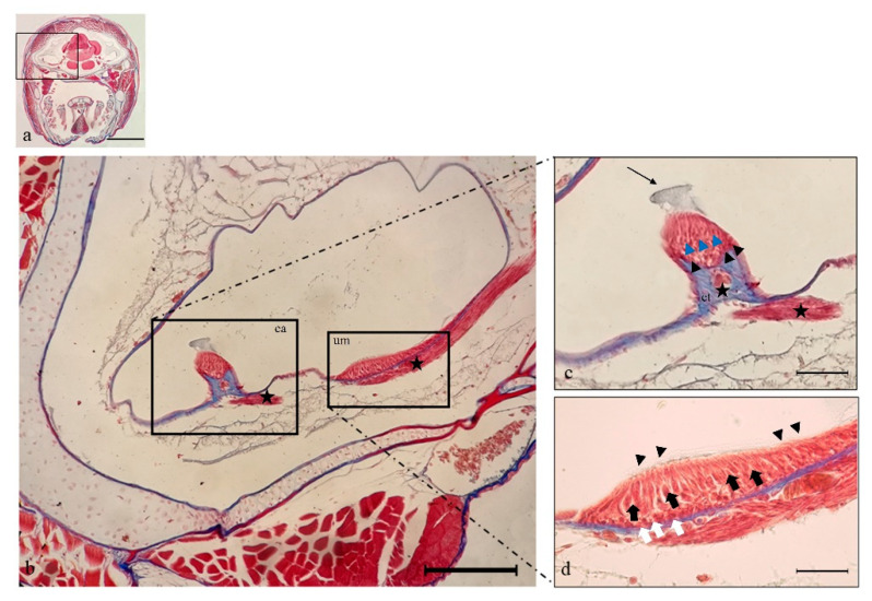
Figure 2.
Light micrographs (Masson Trichrome with Aniline blue staining) of N. guentheri inner ear; transversal view. (a) Head. (b) Semicircular posterior canal of the inner ear, containing the crista ampullaris (ca) and the utricular macula (um). It is possible to observe the crista ampullaris innervation (star) and utricle macula innervation (star). (c) Higher magnification of crista ampullaris in the posterior canal: the connective tissue (ct) supports nerve fibers (star). The black arrowheads indicate the supporting cells, and blue arrowheads indicate the hair cells, the arrow points to the cupula. (d) Higher magnification of utricular macula. The portion of the utricle that forms the macula shows a sort of pouch. The sensory hair cells (black arrows) with numerous stereocilia (arrowheads) are visible. The white arrows indicate the supporting cells. Magnification 4×, scale bar 1 mm (a). Magnification 10×, scale bar 200 µm (b). Magnification 40×, scale bar 50 µm (c,d).

