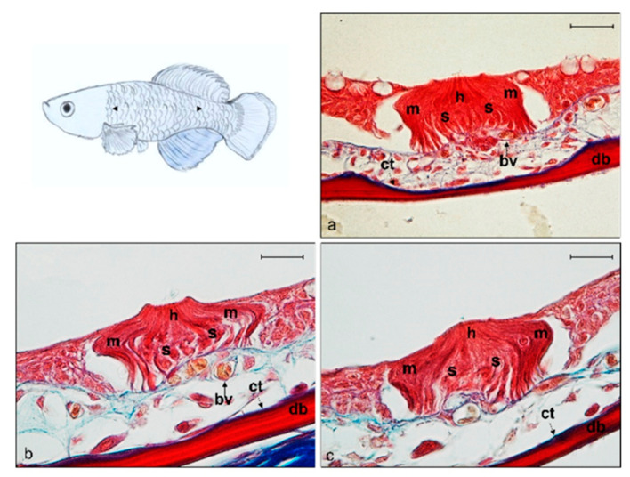
Figure 4.
Light micrographs (Masson Trichrome with blue staining) (a–c) of lateral line system N. guentheri trunk; transversal view. Superficial free neuromast in N. guentheri trunk shows a group of hair sensory cells (h) supported by supporting cells (s) and surrounded laterally by long mantle cells (m). bv (arrow) blood vessels, bm basal membrane, ct (arrow) connective tissue, db dermal bone. Magnification 40×, scale bar 50 µm.

