Figure 8.
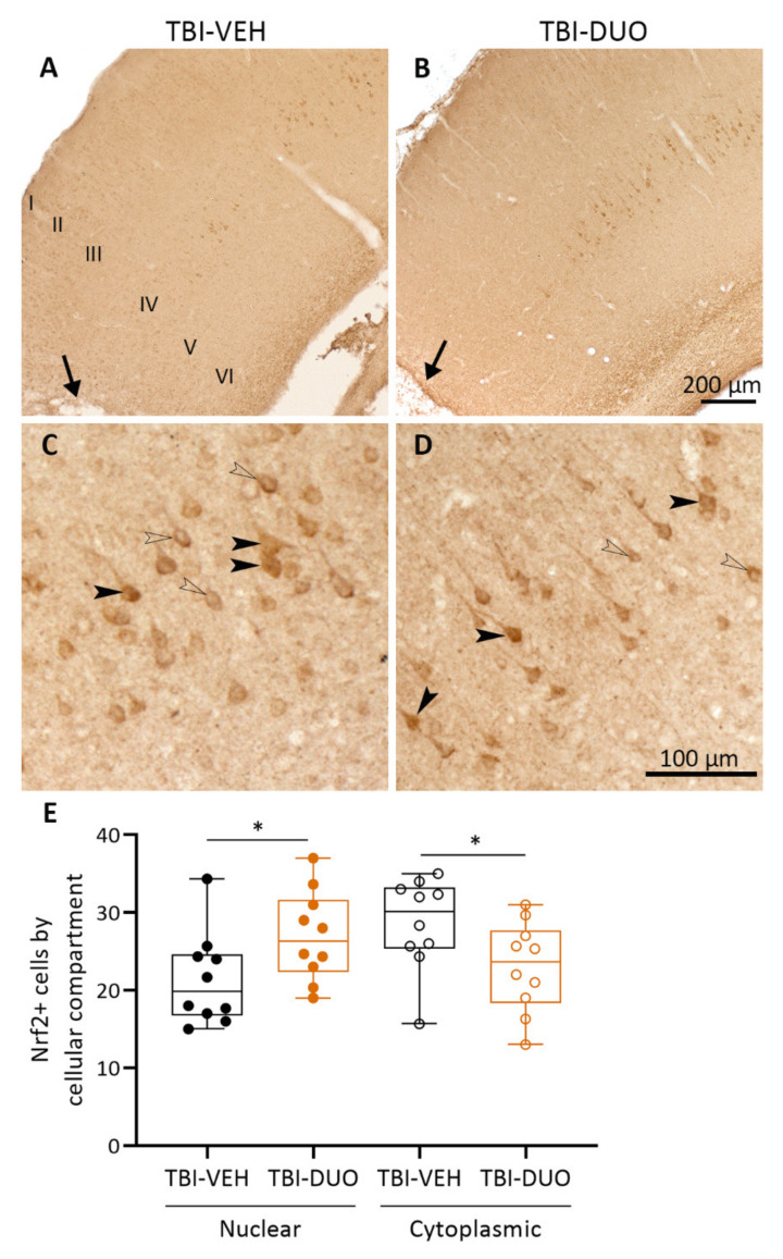
Nrf2+ immunostaining in the ipsilateral cortex. Low magnification photomicrographs showing Nrf2+ immunostaining mainly in cortical layer IV in (A) TBI-VEH and (B) TBI-DUO animals on day (D) 14 post-TBI. Note the lack of immunopositive neurons in a ~1-mm wide sector from the lesion core (lesion core is indicated with an arrow in panels A and B). Cortical layers are labeled with Roman numerals. High magnification photomicrographs showing nuclear (closed arrowhead) and cytoplasmic (open arrowhead) staining of Nrf2 in (C) TBI-VEH and (D) TBI-DUO animals. (E) Quantitative analysis indicated that the duotherapy increased nuclear localization and decreased cytoplasmic localization of Nrf2. Abbreviations: D, day; DUO, duotherapy treated with N-acetylcysteine and sulforaphane; Nrf2, nuclear factor erythroid 2-related factor-2; TBI, traumatic brain injury; VEH, vehicle. Statistical significances: * p < 0.05 Student’s t-test. Data are presented as whisker-plots with mean and minimum/maximum. Scale bar equals 200 µm in panels A–B and 100 µm in panels C–D.

