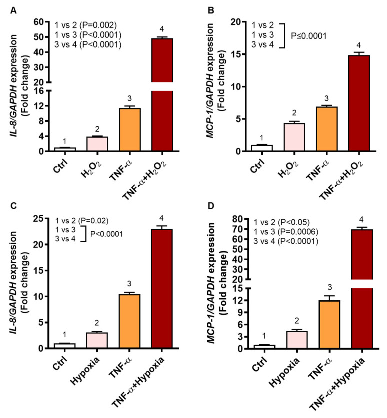
Figure 2.
IL8 and MCP1 gene expression in human monocytic THP-1 cells stimulated with TNF-α and/or oxidative stress. THP-1 cells were stimulated, in triplicate, with TNF-α (10 ng/mL) and/or H2O2 (10 mM) for 24 h while control cells were treated with vehicle only. Likewise, THP-1 cells were also stimulated, in triplicate, with TNF-α under oxidative stress by 1% hypoxia. Total RNA was collected from cells for measuring gene expression of IL8 and MCP1 using qRT-PCR as detailed in methods. One-way ANOVA (Tukey’s multiple comparisons test) was used to calculate group differences and p-values less than 0.05 were considered as significant. The representative data (mean ± SEM) obtained from three independent experiments with similar results show the upregulated transcripts of (A) IL8 and (B) MCP1 in cells co-stimulated with TNF-α and H2O2, compared to TNF-α stimulation alone (p ≤ 0.0001). Similarly, the representative data (mean ± SEM) from three independent experiments with similar results show increased transcripts expression of (C) IL8 and (D) MCP1 in the cells stimulated with TNF-α under 1% hypoxia, compared to TNF-α stimulation under normoxia (20% O2) (p < 0.0001).

