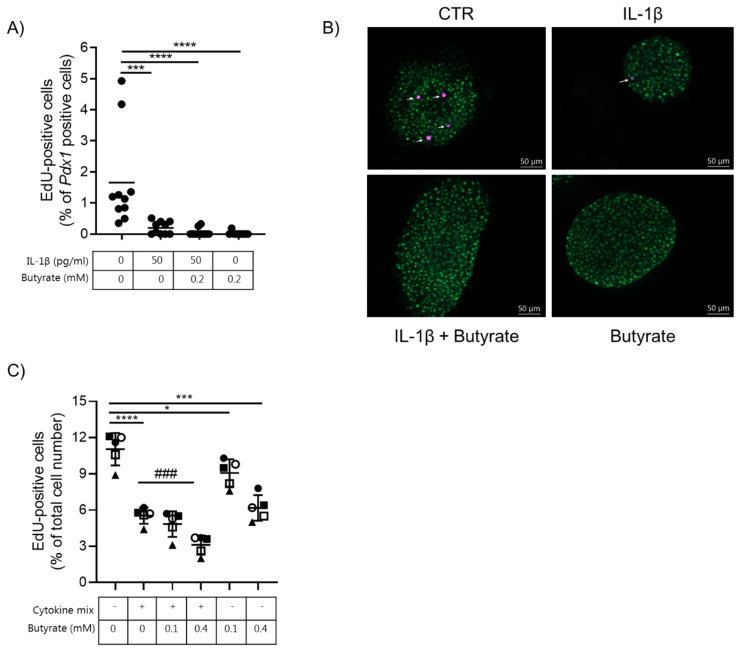
Figure 3.
Butyrate and cytokines reduce mouse islet and human EndoC-βH1 beta cell proliferation. (A) Beta cell proliferation was examined in mouse islets exposed to 50 pg/mL IL-1β and/or 0.2 mM butyrate, or left non-exposed for 10 days. Proliferation was determined by staining for Pdx1 and EdU. Results are shown as percentage proliferating beta cells of total Pdx1 positive cells. Data are shown for n = 10 islets from three independent experiments and analyzed by an unpaired t-test. *** p < 0.001, **** p < 0.0001 vs. control. (B) Immunocytochemical staining of mouse islets. Cells were stained for Pdx1 (green) and EdU (magenta). Data shown are representative. (C) Beta cell proliferation was measured in EndoC-βH1 cells exposed to cytokine mix (500 pg/mL IL-1β + 2 ng/mL INF-γ + 2 ng/mL TNF-α), 0.1 or 0.4 mM butyrate, or left non-exposed for 6.5 days. Proliferation was measured as EdU-positive cells and shown as the percentage proliferating beta cells of the total cell number per well. Data are shown as means ± SD for n = 5 and analyzed by an unpaired t-test * p < 0.05, *** p < 0.001, **** p < 0.0001 vs. control, ### p < 0.001 vs. IL-1β. Each condition was tested in biological triplicate.

