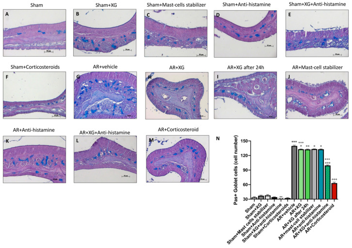
Figure 11.
Effect of XG treatment on PAS staining. An important increase of PAS positive cells was detected in AR-mice (G, histological score N), compared to sham mice (A–F, histological score N). XG treatment (H,I, histological score N) diminished the mucous secretion observed as PAS-positive cell number with equivalent effectiveness to the antihistamine (K, histological score N) and mast-cell stabilizer (J, histological score N) treatments. In addition, co-treatment with XG and antihistamine (L, histological score N) reduced PAS-positive cells, in a comparable way with corticosteroid treatment (M, histological score N). Data are representative of at least three independent experiments. Values are means ± SEM. One-Way ANOVA test. *** p < 0.001 vs. Sham; ° p < 0.05 vs. AR + vehicle; °° p < 0.01 vs. AR + vehicle; °°° p < 0.001 vs. AR + vehicle; +++ p 0.001 vs. AR + Corticosteroid.

