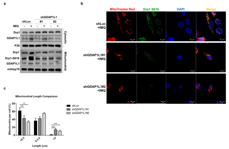
Figure 3.
Loss of GDAP1L1 ameliorated Drp1 phosphorylation and mitochondrial translocation. Control and GDAP1L1-deficient THP1 macrophages were stimulated with IMQ (10 μg/mL) for 2 h. (a) Cells were fractionated into cytosolic and mitochondrial fractions and subjected to immunoblotting using the indicated antibodies. (b) Cells were stained with MitoTracker Red and fixed and immunostained with antibodies against GDAP1L1 and Drp1 S616, followed by confocal microscopy. Nuclei were stained blue using DAPI. (c) Average mitochondrial length (μm) and comparison of mitochondrial length distribution in the indicated groups. Scale bars, 10 μm. All experiments were repeated two or three times with similar results. Significant differences are indicated by ** p < 0.01 and, *** p < 0.001. Data are represented as mean ± SEM.

