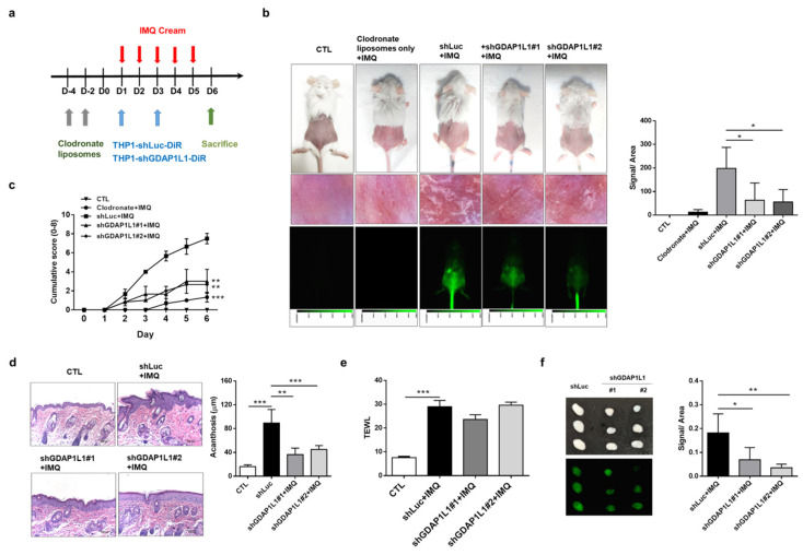
Figure 4.
GDAP1L1 inversely regulated the macrophage numbers in lesional skin and the skin draining lymph node, and the histological changes in IMQ-induced psoriasis-like inflammation. (a) Experimental schedule for DiR-labeled control or GDAP1L1-deficient THP1 macrophage transplantation into the macrophage-depleted psoriasis-like inflammation model. (b) Phenotypical and microscopic images of IMQ-induced macrophage-depleted psoriasis-like inflammation on mouse back skin with and without control or GDAP1L1-deficient THP1 macrophage transplantation. The in vivo biodistribution of DiR-labeled THP1 cells of the skin was monitored using Pearl Impulse (Li-Cor, NE, USA) after intravenous injection in a mouse. In vivo accumulation signal in a mouse, skin injection of DiR-labeled THP1 cells, was quantified in the right panel (n = 6). (c) The cumulative score (erythema plus scaling on a scale from 0 to 4 of each) was depicted (0–8) (n = 6). (d) Histological assessment of back skin by H&E staining and thickness (μm) measured using an image analysis system in the right panel (n = 8). (e) Transepidermal water loss (TEWL) was measured at day 6 (n = 6). (f) At day 6, the lymph node was removed and visualized as shown, and ex vivo near-infrared signal of the organs was quantified (n = 5). All experiments were performed at least three times. * p < 0.05, ** p < 0.01 and *** p < 0.001 when compared to control group. Data are represented as mean ± SEM. Scale bar, 100 μm.

