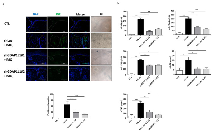
Figure 5.
Loss of GDAP1L1 attenuated inflammation cytokine expression in an IMQ-induced mouse model. (a) Skin cryosections from the DiR-labeled control or GDAP1L1-deficient THP1 macrophage transplantation mice. DiR-labeled infiltrated macrophages (green) were found in the dermis and nearby thickened epidermis, as indicated by arrows. Cell nuclei (blue) were counterstained with DAPI. The dotted lines indicate the border between the epidermis and dermis. Scale bar, 100 μm. Quantitative analyses of DiR-labeled infiltrated macrophages in the skin were counted in high-power fields. (n = 5) is presented in the bottom panel. (b) Production of cytokines as indicated in the skin (n = 5−6) was measured with ELISA. * p < 0.05, ** p < 0.01 and *** p < 0.001 when compared to control group. Data are represented as mean ± SEM of three independent experiments.

