Figure 1.
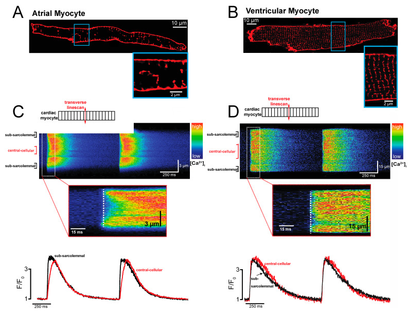
Spatiotemporal Ca2+ release in atrial and ventricular cardiac myocytes. (A) Transverse tubules. Confocal laser scanning image of a freshly isolated living atrial myocyte stained with the membrane dye Di-8-ANEPPS. (B) The same as in (A), but in a freshly isolated living ventricular myocyte. (C) Intracellular Ca2+ release. Shown is an atrial myocyte loaded with the fluorescent Ca2+ indicator Fluo-4. A transverse confocal linescan is used to track the spatio-temporal intracellular Ca2+ release from the subsarcolemmal domain to the center of the cell in an atrial myocyte during external field stimulation. The inset shows the onset of Ca2+ release at the outer cell membrane in the subsarcolemmal space compared to the central cellular domain. Below are the domain-specific Ca2+ transients derived from the confocal images shown in the upper panel. (D) The same as in (C) but for a ventricular myocyte.

