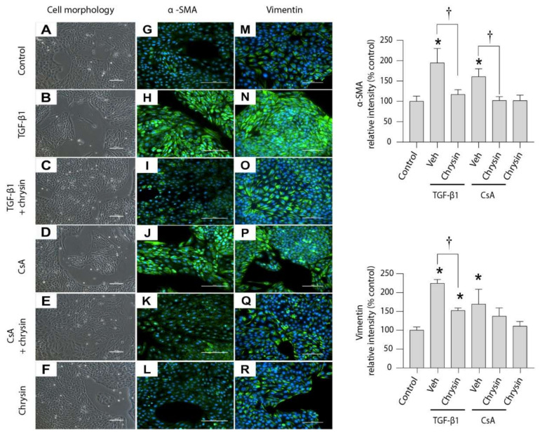
Figure 4.
Effect of chrysin on TGF-β1- or CsA-induced EMT. LLC-PK1 cells were treated for 48 h with 5 ng/mL TGF-β1 or 4.2 µM CsA in the absence of 25 µM chrysin. Morphological changes (A–F) and immunofluorescence (green) of α-SMA (G–L) and vimentin (M–R) were assessed. Micrographs shown are representative of four individual experiments. For immunocytochemistry the nuclei were counterstained with DAPI (blue). The light micrographs were taken at a magnification of 200× (scale bar = 100 µm) and immunofluorescence micrographs were captured at a magnification of 400× (scale bar = 100 µm). Fluorescence for α-SMA and vimentin was quantified and data are represented as percent intensity relative to the control group. Statistical significance was determined by two-way ANOVA followed by Tukey’s multiple comparison test. * indicates p < 0.05 versus control; † indicates p < 0.05 versus chrysin co-treatment.

