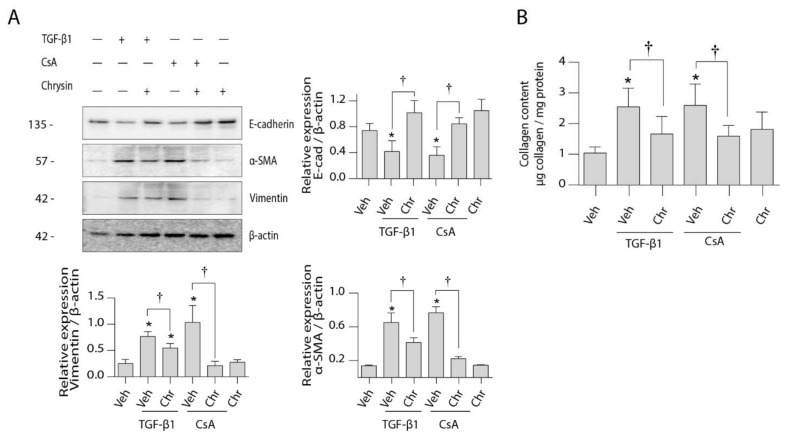
Figure 5.
Effect of chrysin on CsA- or TGF-β1-induced EMT protein expression. LLC-PK1 cells were treated with vehicle (Veh), 4.2 µM CsA, 5 ng/mL TGF-β1 or 25 µM chrysin (Chr) or both for 48 h. (A) Protein expression of epithelial and mesenchymal markers were assayed using the Western blot method. Representative Western blots for E-cadherin, αSMA and vimentin are shown, and data are represented as mean intensity relative to β-actin ± SD of three individual experiments. (B) The amount of collagen deposited in the cell monolayers was assayed by Sircol™ Sirius red assay and normalized to protein content. Data are represented as mean collagen content ± SEM of four individual experiments. Statistical significance was determined by one-way ANOVA followed by Tukey’s multiple comparison test. * indicates p < 0.05 versus control; † indicates p < 0.05 versus chrysin co-treatment.

