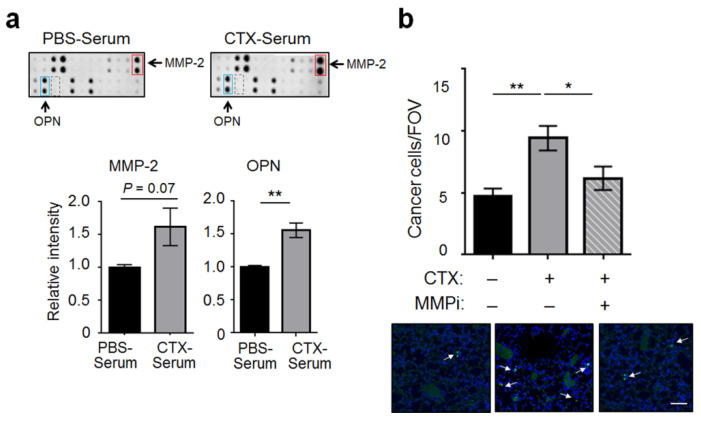
Figure 4.
CTX increased the serum level of MMP-2, which was functionally important for CTX to increase the adhesiveness of vascular walls. (a) Top: A portion of the protein array is shown for MMP-2 (red box), OPN (blue box), and MMP-9 (dashed box). Bottom: Signals of MMP-2 and OPN were normalized as detailed in Materials and Methods (subtract the background and normalized against the positive controls). The normalized signals from the PBS-serum blot were arbitrarily defined as 1. (b) Mice were pre-treated with CTX (+) or vehicle (−), followed by injection of MMP-2/9 inhibitor III (MMPi) (+) or vehicle (−) 24 and 72 h after CTX treatment. Cancer cell number per FOV at 3 h post-injection were analyzed (n = 6–9 from 2 independent experiments). Bottom: Representative images. Arrows indicate cancer cells, which are discrete and bright, in contrast to the background noise of faint and diffuse nature (presumably due to the auto-fluorescence of bronchioles and/or blood vessels). Scale bar, 50 μm. Bars indicate mean ± SEM; one-way ANOVA with post hoc Holm–Šídák correction; * p < 0.05; ** p < 0.01.

