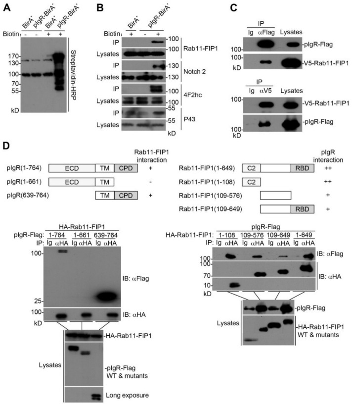
Figure 1.
Rab11−FIP1 interacts with pIgR. (A) HEK293T cells (2 × 10⁶) were transfected with pIgR−BirA* or BirA* plasmids for 24 h. Cells were untreated or treated with 50 µM biotin for 12 h before lysis in the indicated buffer. Biotin−labelled proteins were immunoprecipitated from lysates using streptavidin conjugated agarose beads. Immunoprecipitated proteins were detected by immunoblotting analysis and identified by mass spectrometry. (B) The samples of (A) were respectively detected by immunoblotting analysis with the indicated antibodies. (C) Detection of the interaction between Rab11−FIP1 and pIgR. HEK293T cells (2 × 10⁶) were co−transfected with the indicated plasmids for 24 h. Coimmunoprecipitation and immunoblotting analyses were performed with the indicated antibodies. (D) Domain mapping of the interaction between Rab11−FIP1 and pIgR. HEK293T cells (2 × 10⁶) were co−transfected with the indicated plasmids for 24 h. Coimmunoprecipitation and immunoblotting analyses were performed with the indicated antibodies. ECD: ectodomain; TM: transmembrane domain; CPD: cytoplasmic domain. ++: stronger interaction, +: interaction, −: no interaction. Data of (A–D) are representative of three independent experiments.

