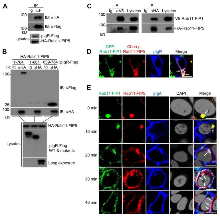
Figure 4.
The Rab11−FIP1, Rab11−FIP5 and pIgR complex facilitates pIgA transcytosis. (A) The interaction between Rab11−FIP5 and pIgR was detected. HEK293T cells (2 × 10⁶) were co−transfected with the indicated plasmids for 24 h. Coimmunoprecipitation and immunoblot analyses were performed with the indicated antibodies. (B) Domain mapping of the interaction between Rab11−FIP5 and pIgR. HEK293T cells (2 × 10⁶) were co−transfected with the indicated plasmids for 24 h. Coimmunoprecipitation and immunoblot analysis were performed with the indicated antibodies. (C) The interaction between Rab11−FIP1 and Rab11−FIP5 was detected. HEK293T cells (2 × 10⁶) were co−transfected with the indicated plasmids for 24 h. Coimmunoprecipitation and immunoblotting analyses were performed with the indicated antibodies. (D) Colocalization of Rab11−FIP1, Rab11−FIP5 and pIgR was detected. Vero−pIgR cells (1 × 10⁵) were co−transfected with GFP−Rab11−FIP1 (0.2 µg) and Cherry−Rab11−FIP5 (0.2 µg) for 24 h. The transfected cells were fixed with 4% paraformaldehyde and stained with the indicated antibodies before observation by confocal microscopy. Scale bar: 10 µm. (E) Colocalization of Rab11−FIP1, Rab11−FIP5 and pIgA was detected during pIgA transcytosis. Vero−pIgR cells (1 × 10⁵) were grown on Transwell for 3 days. An amount of 20 µg pIgA was added or not added to the basal chamber for 10 min at 37 °C and cells were then washed for three times. Subsequently, cells were cultivated at 37 °C and harvested at the indicated time points. Finally, the cells were fixed with 4% paraformaldehyde and stained with the indicated antibodies before observation by confocal microscopy. Scale bar: 10 µm. Data of (A–E) are representative of three independent experiments.

