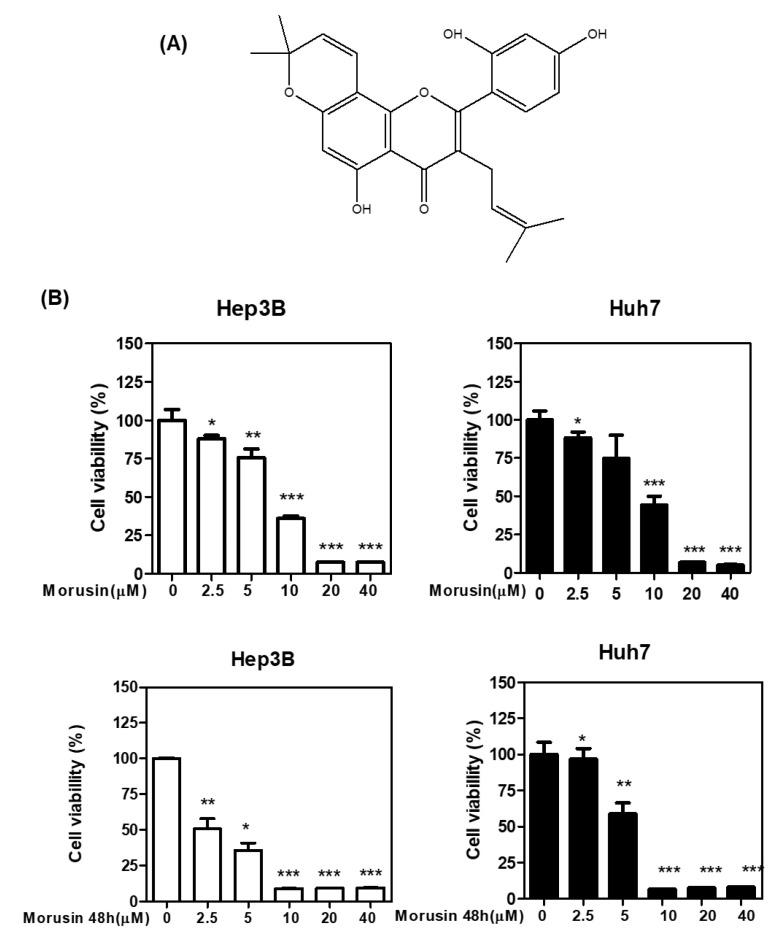
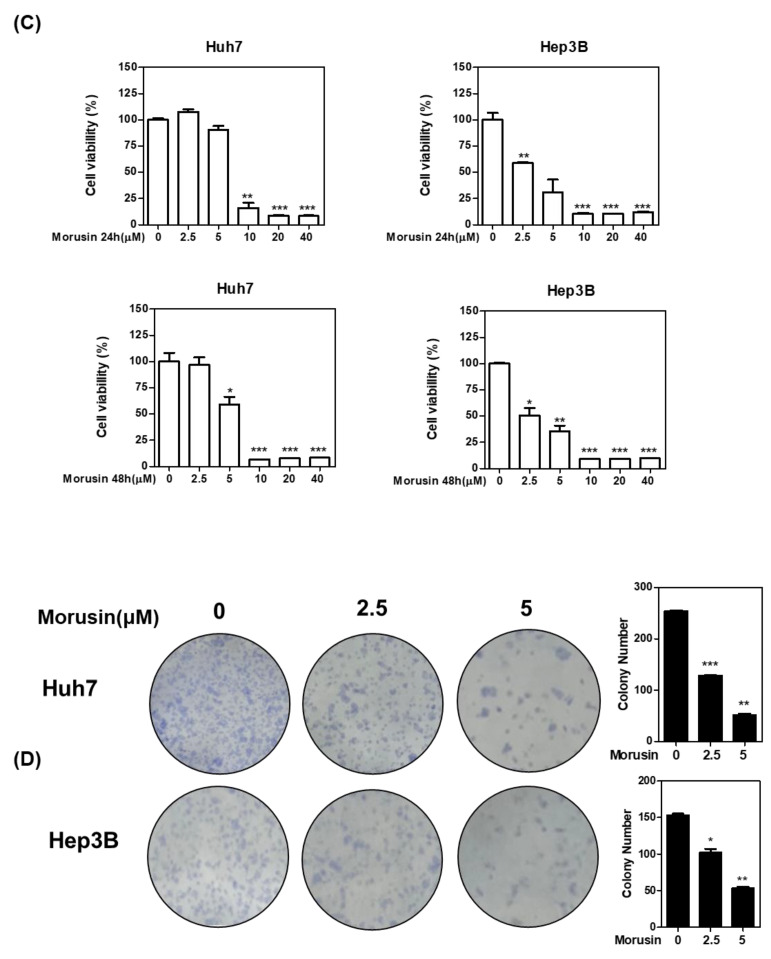
Figure 1.
Chemical structure of Morusin and its effect on cytotoxicity in HCCs. (A) Chemical structure of Morusin (B) Effect of Morusin on the viability of Huh7 and Hep3B cells by MTT assay. Huh7 and Hep3B cells were seeded into 96-well microplates and treated with various concentrations (2.5, 5, 10, 20, and 40 µM) of Morusin for 24 h and 48 h. Cell viability was measured by MTT assay. * p < 0.05, **, p < 0.01, ***, p < 0.001 vs. untreated control. (C) Effect of Morusin on Huh7 and Hep3B cells. Cells were cultured with Morusin (0–40 μM) for 24 h and 48 h and then measured by CCK-8 assay (D) Effect of Morusin on the number of colonies in Hep3B cells by colony formation assay. Huh7 and Hep3B cells were seeded onto 6-well plates for a week. Then the colonies stained with crystal violet were counted. Data represent means ± SD. *, p < 0.05, **, p < 0.01, ***, p < 0.001 vs. untreated control (n = 3).


