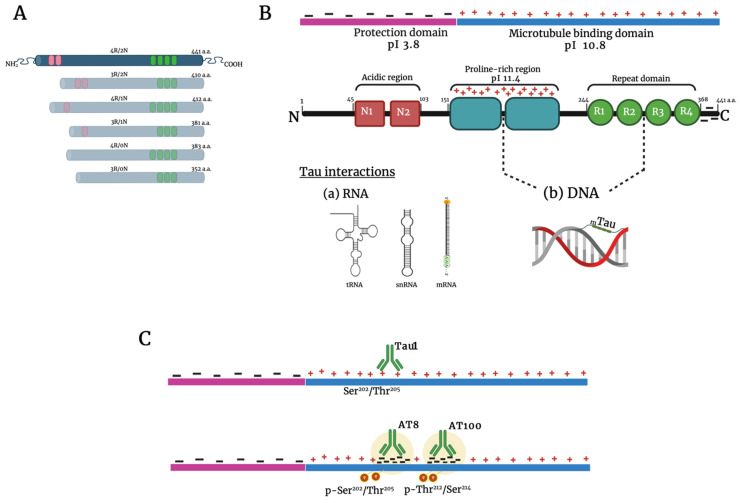
Figure 1.
Tau protein structure and nuclear interactions. Structure of the six tau isoforms expressed in the adult human brain (A). Details of the longest isoform. It contains three major domains: an acidic N-terminal part (red), a proline-rich region (blue), and the basic microtubule-binding domain (green). The electrostatic interactions between domains allow the formation of condensed liquid tau droplets. The dotted line indicates the region where tau interacts with DNA and RNA (B). Upper line: Tau1 antibody recognizes the Ser202/Thr205 site. Lower line: The antibodies AT8 and AT100 bind with the phosphorylated sites Ser202/Thr205and Thr212/Ser214, respectively. Notice the change in the charge of the protein upon phosphorylation (C). Abbreviations: pI, regional isoelectric points; a.a., amino acids. Created with BioRender.com.

