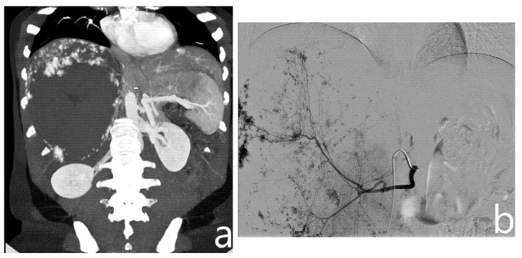
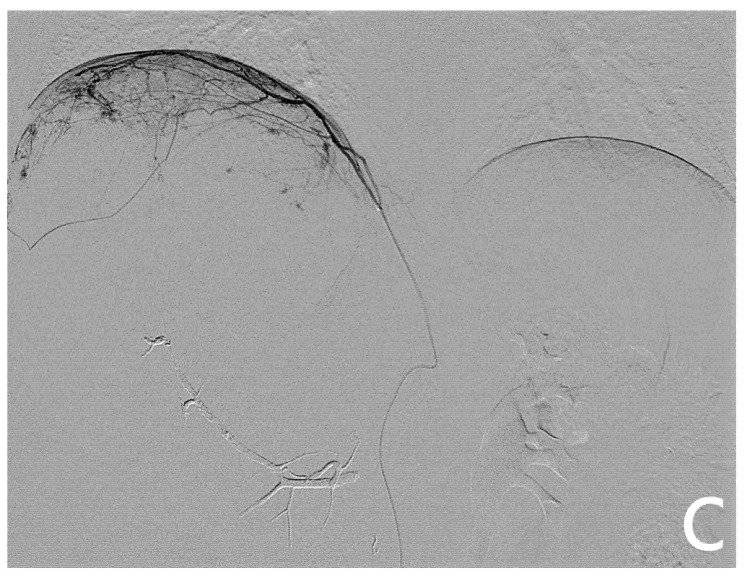
Figure 1.
On preoperative CT (a), a giant hemangioma of the right liver lobe is identified, with evidence of vascular afference from the right phrenic artery (arrow). On angiography, after identification and embolization of the right hepatic artery (b), right phrenic artery is selectively catheterized and embolized with microparticles (c).


