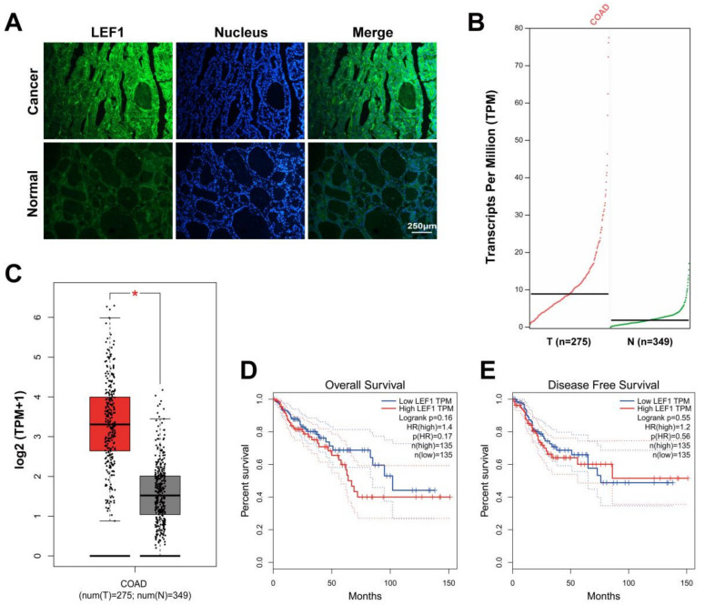
Figure 1.
LEF1 was overexpressed in colonic adenocarcinoma tissues and reinforced the progression of colonic adenocarcinoma. (A): The expression level of LEF1 in colonic adenocarcinoma tissues was analyzed by IF staining (scale bar = 250 μm). (B,C): The LEF1 mRNA expression level in tumor tissues and paired normal tissues (* p < 0.05). (D,E): Kaplan-Meier curves showing the 10-year overall survival rate and disease free survival rate in patients with high LEF1 expression (n = 135) and low LEF1 expression (n = 135). The data were analyzed by one-way ANOVA.

