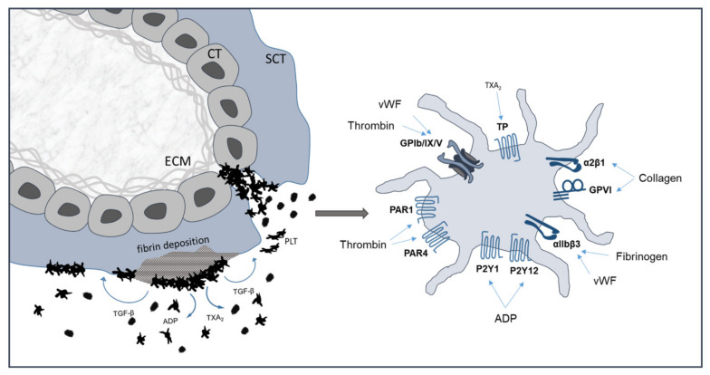
Figure 3.
Platelet activation at the maternal-fetal interface. Scheme of a first trimester placental villus shows adherent platelets on fibrin deposition and on extracellular matrix on areas of disrupted villous trophoblasts. Activated platelet show relevant surface receptors. CT: cytotrophoblast; SCT: syncytiotrophoblast; ECM: extracellular matrix; PLT: platelet; TXA2: thromboxane A2; ADP: adenosine diphosphate; TGF-β: transforming growth factor beta; vWF: von Willebrand factor; TP: thromboxane receptor.

