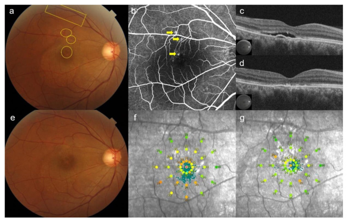
Figure 6.
Case 2: A 45-year-old man presented with an 8-month history of blurred vision in the right eye. After the 6-week observation period, subretinal fluid was still observed at the macula on color fundus photography. The area of 13 test spots (yellow rectangular) and 15 rescue treatment spots (yellow circles) were invisible (a). Three focal leakages (yellow arrows) were shown on fundus fluorescent angiography (b). Subretinal fluid and pigment epithelial detachment was observed on optical coherence tomography (OCT) before rescue with selective retina therapy (SRT) (c). At 6-weeks post-SRT, subretinal fluid was completely resolved on OCT (d). No SRT spots were visible on fundus photography (e). Mean retinal sensitivity (20 dB) on microperimetry at baseline (f) was increased to (22 dB) on microperimetry (g) at 6-weeks post-treatment.

