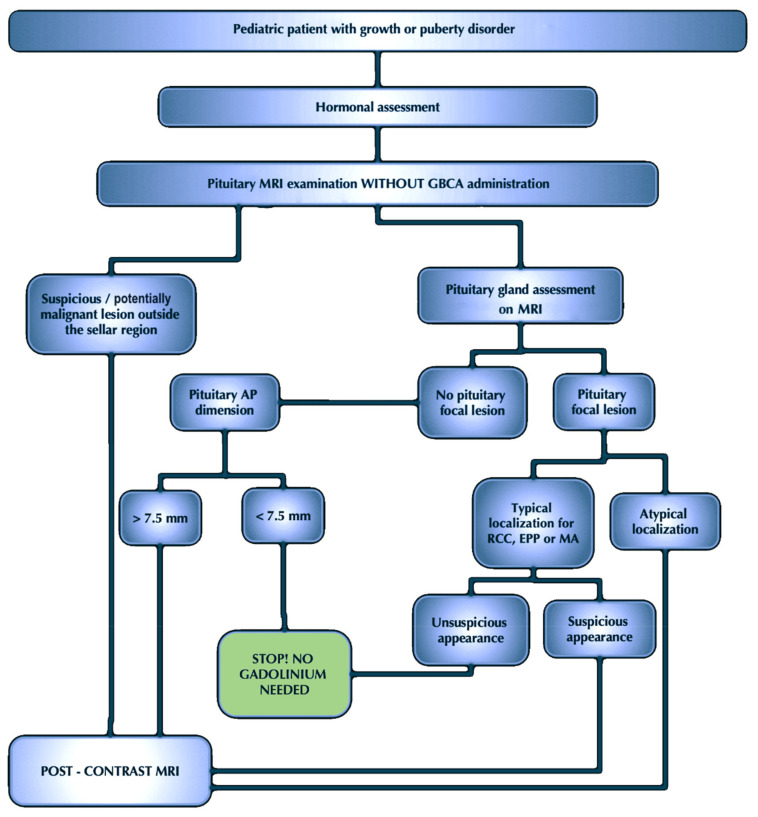
Figure 6.
The practical approach for assessment of the pituitary MRI in children with growth or puberty disorders before hormonal therapy. In the new imaging diagnosis algorithm of children with GPDs proposed by us, after endocrinological assessment, patients would have a pituitary MRI examination without GBCA. The results of our study show unequivocally that in most cases, the analysis of native sequences is sufficient to assess the gland to the extent that it allows the patient to qualify for substitution treatment. If the native pituitary MR examination reveals no focal lesion in the parasellar region, and the pituitary AP dimension does not exceed 7.5 mm, the statistical probability of detecting a focal lesion in post-contrast sequences is so small that it is possible to omit gadolinium administration. However, if the AP dimension exceeds 7.5 mm, the examination should be extended by post-contrast sequences. In turn, if a focal lesion in the native MRI is visible, its location and morphology should be carefully assessed. If the lesion location is typical for RCC, EPP, or MA, and the morphology of the focal lesion does not raise suspicions, administration of the contrast agent should also be omitted. However, if the focal lesion has an atypical location, or the location corresponds to RCC, EPP, or MA but its morphology is of concern, in these cases, the pituitary MRI should be extended by post-contrast sequences. Of course, if a suspicious or potentially malignant focal lesion is observed beyond the parasellar region, not only should gadolinium be administered, but the scope of the examination should also be extended, and the entire brain should be visualized.

