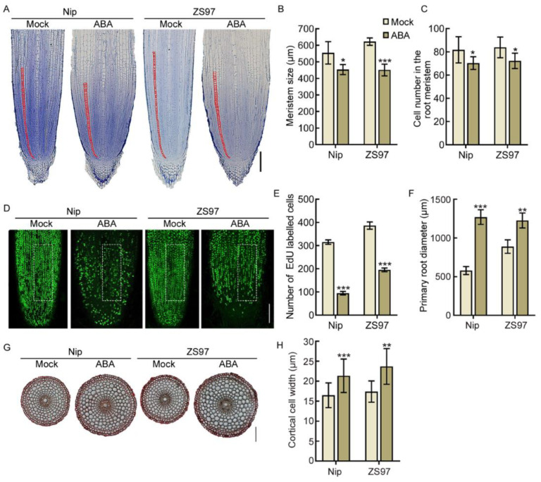
Figure 4.
ABA inhibits cell proliferation and promotes cortical cell expansion in the root meristem. (A) Longitudinal sections of primary root tips (lower) of 4-day-old Nip and ZS97 seedlings treated with 0 μM (Mock) and 1 μM ABA. Red lines delimit the meristem size (i.e., the distance between the quiescent center and the transition zone). Bar = 100 μm. (B,C) Length of root meristematic zone (B) and cortical cell number in the root meristem zone (C) of the corresponding seedlings indicated in (A). (D) EdU-labelled cells in the root meristem of 4-day-old Nip and ZS97 seedlings treated with 0 μM (Mock) and 1 μM ABA. Bar = 100 μm. (E) Number of EdU-labelled cells in (D). Quantification was performed in the unit area (white box) of the root tip. (F) Primary root diameter of the corresponding seedlings indicated in (A). (G) Cross-sections of primary root tips (lower) of 4-day-old Nip and ZS97 seedlings with or without 1 μM ABA treatment. Bar = 100 μm. (H) Diameter of cortical cells in (G). In (B,C,E,F,H), data are means ± SD (n ≥ 10 independent seedlings). The asterisks indicate significant differences compared to mock (* p < 0.05, ** p < 0.01, *** p < 0.001, Student’s t-test).

