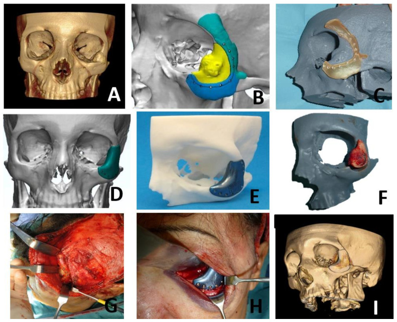
Figure 5.
A 55-year-old woman, smoker, with a history of multiple sclerosis, reported a tumor of progressive growth and pain in the left zygomatic region. After study by CT, MRI and arteriography, the patient was diagnosed with intraosseous vascular malformation in the zygomatic bone (A). Virtual surgical planning was performed for resection and reconstruction with a titanium PSI. Cutting guides were designed for resection (B,C). A customized titanium PSI was specifically designed for the reconstruction (D,E). A hemicoronal and subciliary approach were performed and cutting guides were used to perform the resection (F–H). Once the lesion was removed, the titanium PSI was placed with an excellent fit (H). The postoperative CT scan showed a correct placement of the titanium PSI with an optimal aesthetic result (I).

