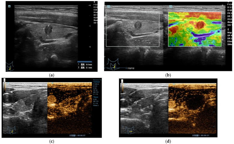
Figure 1.
(a) At B-mode US, the shape of the lesion appeared taller-than-wide, hypoechoic with regular margins without internal microcalcifications (EU-TIRADS 5); (b) At qualitative USE evaluation, the lesion appeared stiff (completely red in the color box); (c) At CEUS evaluation, the lesion appeared richly vascularized similar to surrounding thyroid parenchyma without strong wash-out. At FNAC the lesion was classified as Tir 5. (d) After Radiofrequency Ablation, the lesion and surrounding parenchyma does not show enhancement in the CEUS mode.

