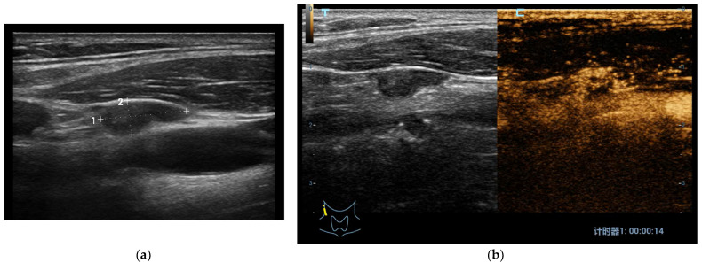
Figure 2.
(a) At B-mode US in Patient with papillary thyroid carcinoma, a laterocervical node showed irregular shape, hypoechoic aspect and regular margins with no internal microcalcifications or cystic changes (low metastatic risk); (b) At CEUS, the laterocervical node presented rich heterogeneous and centripetal vascularization (high metastatic risk). The histological examination confirmed that it was a metastatic node of a papillary thyroid cancer.

