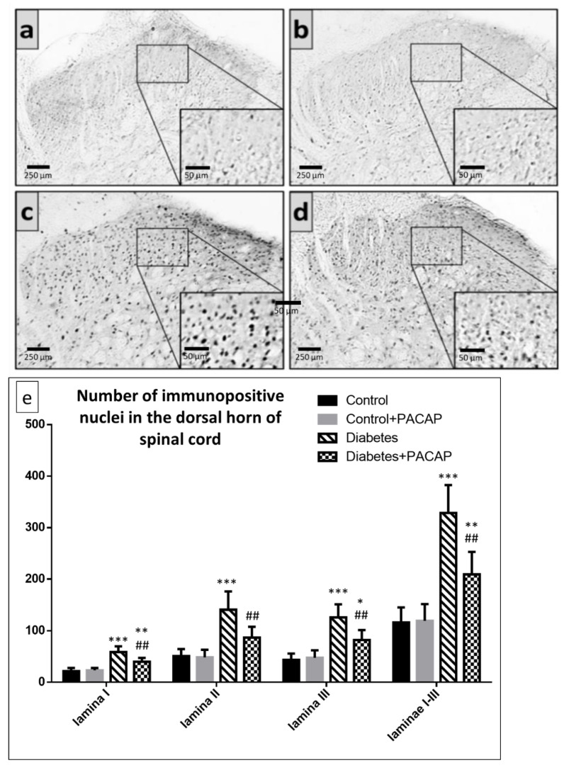
Figure 3.
Representative images of FosB immunohistochemistry in the spinal dorsal horn of segments L4–L5 in (a) vehicle-treated control, (b) PACAP-treated control, (c) vehicle-treated diabetic, and (d) PACAP-treated diabetic groups. Histograms (e) show the number of FosB immunoreactive nuclei in laminae I-III of the spinal dorsal horn of segments L4–L5. Data are means ± SEM of n = 5/6 rats/group.* p < 0.05, ** p < 0.01, *** p < 0.001 vs. vehicle-treated control, ## p < 0.01 vs. vehicle-treated diabetic group.

