FIG. 1.
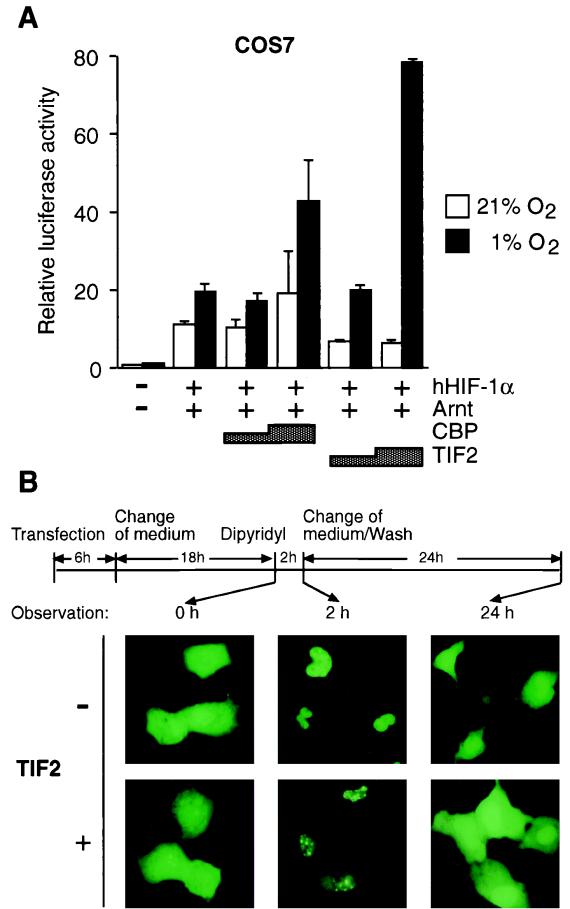
TIF2 enhances the HIF-1α-mediated transactivation function and interacts with HIF-1α in vivo. (A) COS7 cells were transiently cotransfected as described in Materials and Methods with 0.5 μg of pT81/HRE-luc reporter construct, hHIF1-α expression vector (pCMV4/HIF-1α; 0.2 μg), Arnt expression vector (pCMV4/Arnt; 0.2 μg), and 0.75 to 1.5 μg of either TIF2 (pSG5/TIF2) or CBP (Rc/RSV-CBP-HA), as indicated. The total amount of DNA was kept constant by the addition of parental pCMV4 when appropriate. Following transfection cells were incubated either under normoxic (21% O2) or hypoxic (1% O2) conditions. Luciferase activities were normalized for transfection efficiency by cotransfection of alkaline phosphatase-expressing pRSV-AF. Data are presented as luciferase activity relative to cells transfected with pCMV4 and reporter gene only and incubated at normoxia. Values represent the mean ± SE of two independent experiments. (B) Intranuclear redistribution of the GFP-HIF-1α fusion protein in the presence of TIF2. GFP–HIF-1α was transfected into COS7 cells in the absence or presence of exogenous TIF2 (pSG5/TIF2). After 24 h of expression, either 100 μM 2,2′-dipyridyl or vehicle (H2O) was added to the culture medium and incubated for 2 h before observation. Subsequently, 2,2′-dipyridyl was withdrawn by washing the cells and changing the medium, and the cells were thereafter incubated for an additional 24 h. Cells were observed microscopically at different time points as indicated by arrows. Photographs were taken with a Zeiss fluorescence microscope.

