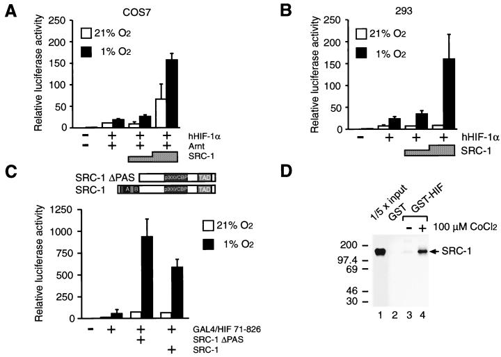
FIG. 2.
SRC-1 stimulates HIF-1α activity in a hypoxia-dependent manner and interacts in vitro with HIF-1α. COS7 (A) and human embryonic kidney 293 (B) cells were cotransfected with pT81/HRE-luc (0.5 μg), 0.2 μg of pCMV4/HIF-1α (hHIF1-α), 0.2 μg of pCMV4/Arnt (Arnt), and 0.75 to 1.5 μg of SRC-1 (pSG5/SRC-1), as indicated. Six hours after transfection, cells were exposed to either 21 or 1% O2 for 36 h before harvest. Luciferase values are presented as relative luciferase activity as described in the legend to Fig. 1. The results of three independent experiments performed in duplicate ± SE are shown. (C) The SRC-1 PAS domain is not required for functional interaction with HIF-1α. (Top) Schematic representation of full-length SRC-1 and SRC-1ΔPAS. (Bottom) pGAL4/HIF 71-826 was cotransfected into COS7 cells together with a reporter plasmid expressing the luciferase gene driven by the thymidine kinase minimal promoter under the control of five copies of GAL4 binding sites and 1.5 μg of an expression vector encoding either full-length SRC-1 or a deletion mutant of SRC-1, SRC-1ΔPAS, lacking the PAS domain. Cells were exposed to 21 or 1% O2 for 36 h before harvest and reporter gene assays. Luciferase values are presented as relative luciferase activity as described in the legend to Fig. 1. The results of two independent experiments performed in duplicate ± SE are shown. (D) In vitro interaction between SRC-1 and HIF-1α. COS7 cells were transfected with 10 μg of the expression plasmid pGST-HIF-1α (GST-HIF) or empty GST expression vector (GST). Cells were exposed to either 100 μM CoCl2 or vehicle (H2O) for 24 h. Cell extracts were prepared and incubated with [35S]methionine-labeled in vitro-translated SRC-1 protein. The complexes were immobilized on glutathione-agarose beads for 2 h and eluted with the sample buffer by boiling. The eluted material was analyzed by SDS-PAGE (5% gel) and visualized by fluorography. Lane 1 represents one-fifth of the amount of [35S]methionine-labeled SRC-1 used in the binding reactions. Positions of molecular mass markers are shown on the left in kilodaltons.

