FIG. 6.
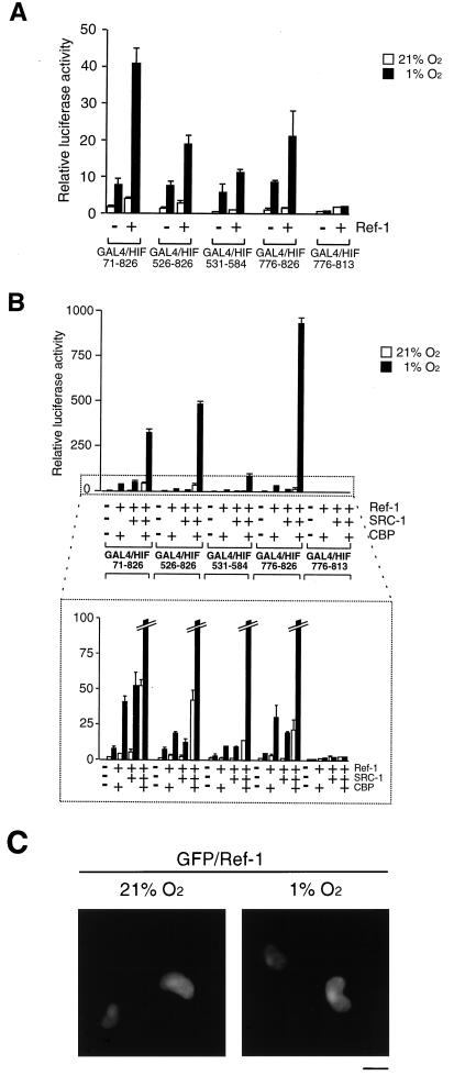
Ref-1 enhances HIF-1α function. (A) COS7 cells were cotransfected with different GAL4–HIF-1α fusion constructs together with a GAL4-responsive reporter plasmid in the absence or presence of 1.5 μg of Ref-1 (pCMV5/Ref-1), as indicated. Cells were exposed to 21 or 1% O2 before harvest. After normalization for transfection efficiency using alkaline phosphatase activity, reporter gene activities were expressed as relative to that of GAL4 in normoxia. The results of two independent experiments performed in duplicate ± SE are shown. (B) Ref-1 potentiates CBP and SRC-1 activation of HIF-1α. The same GAL4– HIF-1α fusion proteins as shown in panel A were cotransfected into COS7 cells together with a GAL4-responsive reporter plasmid in the absence or presence of different combinations of Ref-1 (0.75 μg), CBP (0.75 μg), and/or SRC-1 (0.75 μg) expression vector, as indicated. The bottom panel shows an enlargement of the area marked with dots. (C) Effect of hypoxia treatment on subcellular localization of Ref-1. COS7 cells grown on coverslips were transiently transfected with 3 μg of pGFP/Ref-1. After 6 h of incubation, the medium was changed to fresh DMEM supplemented with 10% FCS and incubated for 24 h. Cells were then exposed to 21 or 1% O2 for 4 h. After being washed three times with PBS, cells were fixed with 4% paraformaldehyde in PBS for 2 h at room temperature, subsequently washed three times with PBS, and mounted. Cells were observed with a fluorescence microscope. Representative cells are shown. Bar = 10 μm.

