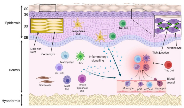
Figure 1.
Physical and immune skin barrier in normal conditions. Densely packed corneocytes, surrounded by a lipidrich extracellular matrix (ECM) in the stratum corneum (SC), prevent passage of water and pathogens. Tight junctions between keratinocytes in the stratum granulosum (SG) prevent paracellular molecular passage. The stratum spinosum (SS) and stratum basale (SB) are deeper and are the initiation site of programmed keratinocyte proliferation and differentiation. Langerhans cells and some T resident memory (Trm) cells are located in the epidermis, while macrophages, conventional dendritic cells (cDCs), mast cells, innate lymphoid cells, γδ T cells and regulatory T cells (Tregs) are located in the uninflamed dermis. In response to pathogens, injury or allergens, these cells are activated. A number of different cytokines and chemokines may be released to compel the infiltration of inflammatory cells, such as neutrophils, monocytes, plasmacytoid dendritic cells (pDCs) and effector T cells (effT) from blood vessels. Collectively, they remove the invaded pathogens and clear cell debris. Regulatory cells resolve inflammation, and the skin barrier is maintained.

