Figure 1.

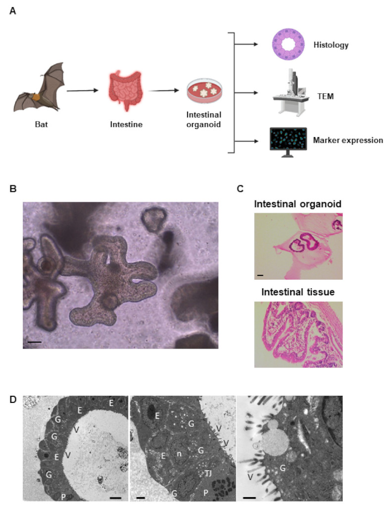
Generation of primary rousette bat intestinal organoids. Experimental schema of establishment and analysis of Rousette bat intestinal organoids (A). Bat intestinal tissues were isolated and cultured for generating their organoids. Then, each tissue-derived organoid was used for the analysis of histology, microstructure by transmission electron microscopy (TEM), and marker expression. Representative phase-contrast images of the organoids (passage 8 at day 10). Scale bar: 200 µm (B). Hematoxylin and eosin-stained bat organoids and original tissues. Scale bar: 100 µm (C). TEM photomicrographs of organoids (D). The absorptive epithelial cells (E), nucleus (n), microvilli (V), tight junction (TJ), goblet cells (G), and Paneth cells (P) are shown. Scale bar: 8 µm, 2 µm, 600 nm from left to right panel, respectively.
