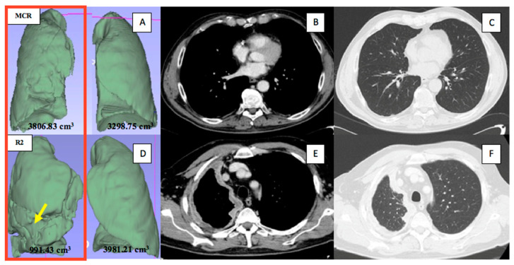
Figure 3.
Representative radiological measurements. Upper panels, a patient with a right-sided MPM affected by the lowest disease burden, who underwent macroscopic complete resection: the aerated ipsilateral and contralateral lung volumes (A) and the pleural thickness (too small to measure) in the axial CT images on the portal venous phase (B) and the lung window (C), at the medium level. Lower panels, a patient with a right-sided MPM affected by the highest disease burden (the arrow indicated the furrows on the three-dimensional lung reconstruction caused by the pleural disease), who underwent R2 resection: the aerated ipsilateral and contralateral lung volumes (D) and the pleural thickness in the axial CT images on the portal venous phase (E) and the lung window (F), at the upper level.

