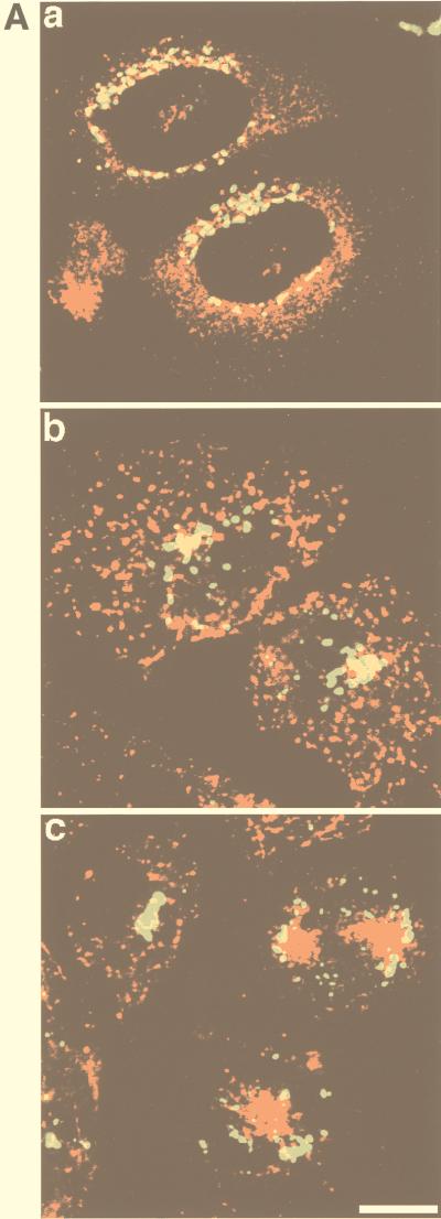
FIG. 6.
Distribution of epitope-tagged sialyltransferase in 3T3-L1 adipocytes and CHO cells. (A) CHO cells stably transfected with GLUT4 or with the human TfR were transiently transfected with VSV-G-tagged sialyltransferase cDNA. GLUT4-sialyltransferase-expressing cells were double labelled with antibodies specific for each of these proteins and visualized using Texas red-conjugated anti-rabbit (GLUT4) or FITC-conjugated anti-mouse (sialyltransferase) secondary antibodies (image a), and TfR-sialyltransferase-expressing cells were incubated with Texas red-conjugated Tf for 15 min (image b) or 60 min (image c) and fixed and labelled with anti-VSV-G antibodies, followed by FITC-conjugated anti-mouse antibodies. Confocal images were obtained with a Bio-Rad MRC-600 laser confocal imaging system. Bar = 10 μm. (B) Membranes from either CHO cells or 3T3-L1 adipocytes, expressing VSV-G-tagged sialyltransferase, were analyzed by iodixanol gradient sedimentation. Gradient fractions were immunoblotted with an anti-VSV-G antibody to detect sialyltransferase. Numbers represent different fractions.


