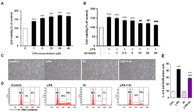
Figure 3.
Ki16425 decreases lysophosphatidic acid (LPA)-induced HaCaT cell proliferation. HaCaT cells were seeded and starved in serum-free media containing 0.1% fatty-acid-free bovine serum albumin (FAF-BSA) for 12 to 16 h. (A) The cells were treated with 1, 5, 10, 20, and 40 μM LPA for 24 h, and cell viability was analyzed by CCK8 assay. (B) The cells were treated with or without 10 μM LPA and indicated ki16425 concentration for 24 h, and cell viability was examined by CCK8 assay. The results are presented as a percentage of control. (C–E) The cells were treated with 10 μM LPA in the presence or absence of 10 μM ki16425 for 24 h. (C) The cell morphology was observed using light microscopy. Original magnification, 100×. (D) Cell cycle phase distribution was analyzed using flow cytometry after staining with propidium iodide for measurement of the cellular DNA content. The values in the representative flow cytometry histograms indicate the percentages of cells in G0/G1, S, and G2/M phases of the cell cycle, in that order. (E) The percentage of cells in the S and G2/M phase was calculated as the sum of each phase from (D). The data are represented as the mean ± standard error of the mean (SEM) of results obtained from three independent experiments. *** p < 0.005 vs. Control; ## p < 0.01, ### p < 0.005 vs. LPA.

