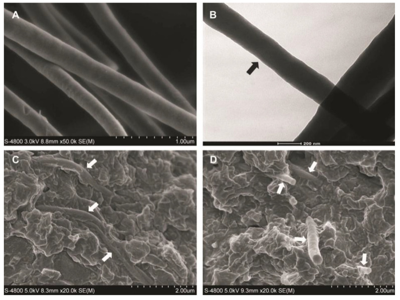
Figure 13.
Morphology of electrospun ZY@S nanofibers: (A) SEM image (fiber diameter: 260 ± 39 nm) and (B) TEM image showing the core-sheath structure. Morphology of the composite fracture surface with (C) 2.5 wt% ZY@S nanofibers and (D) 5.0 wt% ZY@S nanofibers.Images from [98].

