Figure 15.
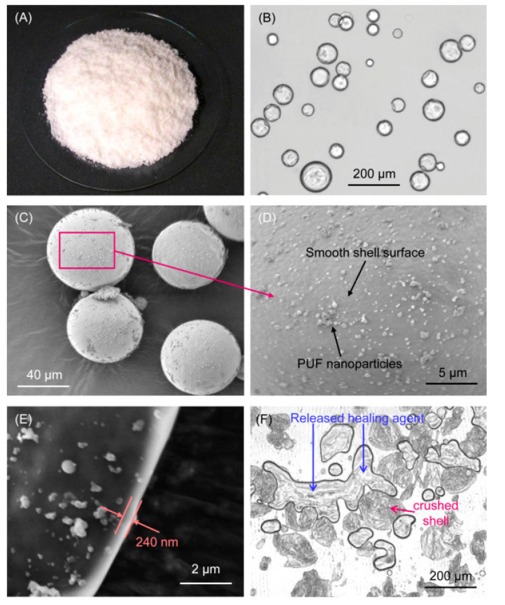
Microcapsules were prepared with a polymerizable TEGDMA–DHEPT healing liquid inside PUF shells. (A) Photo showing a pile of synthesized microcapsules. (B) Transmitting optical image showing the shell structure as a dark ring. (C) SEM image of typical microcapsules. (D) High-magnification SEM image of the shell surface showing nanoparticle deposits on an otherwise smooth shell surface. (E) High-magnification SEM image indicating the shell thickness. (F) Optical image of crushed microcapsules showing the released healing liquid films. Images from [111].

