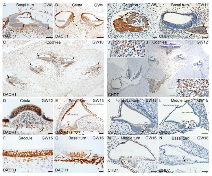
Figure 9.
Immunostaining for DACH1 and CHD7 in human inner ear fetal tissue. (A) Basal turn of cochlea at GW8. Nuclei of future marginal cells (arrow) in stria vascularis are immuno+ for DACH1. Additionally, mesenchyme cells show strong nuclear staining. (B) Cristae ampullaris at GW9. The HCs of lateral and antral crista exhibit strong immunoreactivity in sensory epithelium. Single layered cuboidal epithelial cells of membranous labyrinth are immuno+. Few immuno+ cells observed in the mesenchyme as well. (C) Mid-modiolar plane of cochlea at GW10. Strong immunostaining present in mesenchyme underlying sensory epithelium. Few cells of spiral ganglion exhibit immunoreactivity for DACH1. Future stria vascularis (arrows) is immuno+ for this marker. (D) Crista ampullaris at GW12. Immunoreaction is present in vestibular HCs and supporting cells. Non-myelinating Schwann cells (arrow) underlying sensory epithelium appear immuno+. Transitional cells (TC) are devoid of immunoreactivity. The dark cells (DC) appear immuno+. (E) Cochlear basal turn at GW13. Stria vascularis and Reissner’s membrane immuno+. In organ of Corti (OC), HCs and supporting cells are immuno+. Cells beneath the basilar membrane forming the future tympanic cover layer (TCL) cells are immuno+. (F) Sensory epithelia of saccule at GW15. The HCs and supporting cells show a strong immunoreactivity for DACH1. (G) Cochlea basal turn at GW16. Outer (OHC), inner hair cells (IHC) and supporting cells are immuno+ for DACH1. Tympanic cover layer (TCL) cells exhibit strong immunoreactivity. (H) CHD7 staining in the vestibular ganglion (VG) gestational week (GW) 09. Strong staining in the nerve fibers (arrow). Cytoplasm of the vestibular ganglion cells are also immuno+. Satellite glial cells surrounding the vestibular ganglions are devoid of immunoreactivity. (I) Basal turn of cochlea at GW11. Immunoreactivity is present in the nerve fibers (arrows) of the spiral ganglion (SPG) at this stage. (J) Overview of the temporal bone at GW12. High immunoreaction observed in the nerve fibers and spiral ganglion at the apical turn (AT) and middle turn (MT). Lowered signal in the basal turn (BT). In the vestibular ganglion, a strong immunostaining is evident at this stage. In the saccule (S) and utricle (U), immunoreactivity is absent. (J’) Insert with higher magnification from the vestibular ganglion where the staining of the nerve fibers is evident. (J’’) Insert with higher magnification from the spiral ganglion (SPG). Strong immunostaining observed in the nerve fibers. (K) Basal turn of the cochlea at GW13. CHD7 are penetrating the organ of Corti (arrow). Outer hair cells (OHC). (L) Middle turn of cochlea at GW15. Only single nerve fibers staining observed at the base of the outer hair cells (OHC). Inner hair cell (IHC) (arrow). (M) Middle turn of cochlea at GW16. The tunnel of organ of Corti is already open. We can recognize the nerve fibers staining (arrow). (N) Basal turn of cochlea at GW18. CHD7 staining is penetrating the IHCs (arrow). Organ of Corti (OC). Scale bars: 200 μm (C,J); 100 µm (A,B,F,G); 50 µm (I,K–N); 20 µm (D,E,H,J’,J’’).

