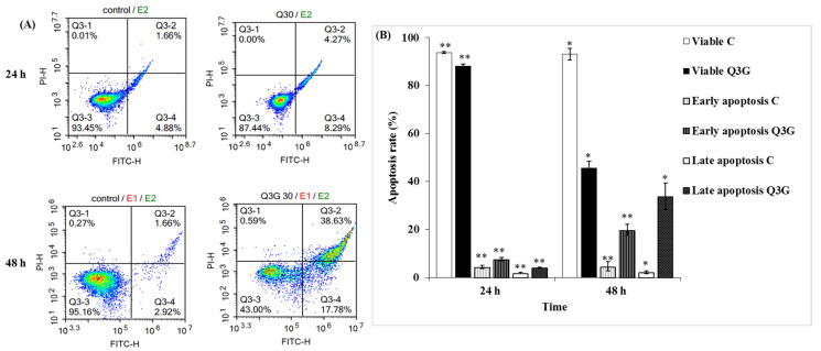
Figure 4.
Q3G induces time-dependent apoptosis in Hela cells. (A) PI and FITC-Annexin V co-staining identified cell death of Hela cells with or without 30 µg Q3G treatment analyzed by flow cytometry. (B) Graph shows the percentages of apoptotic cells. Experiments were performed in triplicate, and the results were expressed as mean ± SD. C: Control, DMSO: dimethyl sulfoxide, PI: propidium iodide, FITC: fluorescein isothiocyanate, Q3G: Quercetin 3-β-D-glucoside. Significantly different from control group, * p < 0.05, ** p < 0.01.

