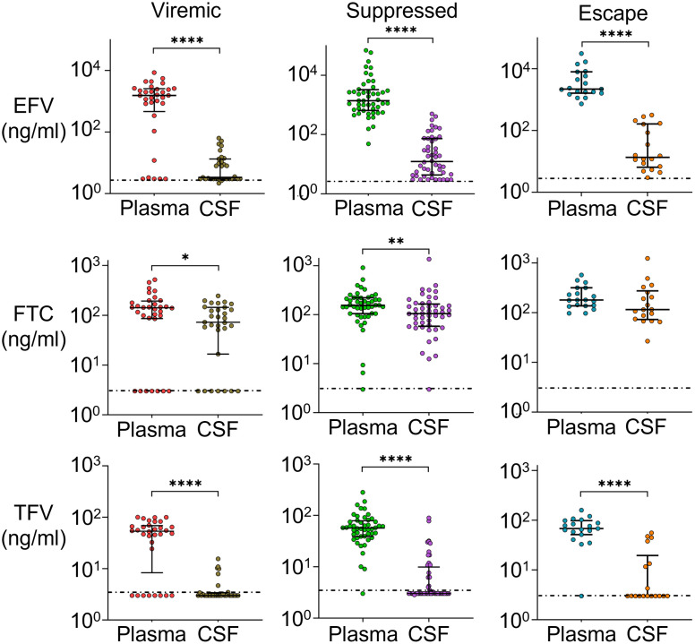
Fig 4. Sharp drop in EFV levels between plasma and CSF.
Medians and IQR of EFV, FTC, and TFV concentrations in the plasma versus CSF in participants who were viremic on ART (plasma n = 32, CSF n = 31), suppressed (plasma and CSF n = 48), or showed CSF escape (plasma n = 19, CSF n = 18). Horizontal dotted line indicates limit of detection (3 ng/mL). p-values are: *< 0.05; **< 0.01; ****< 0.0001 using the Mann-Whitney U test.

