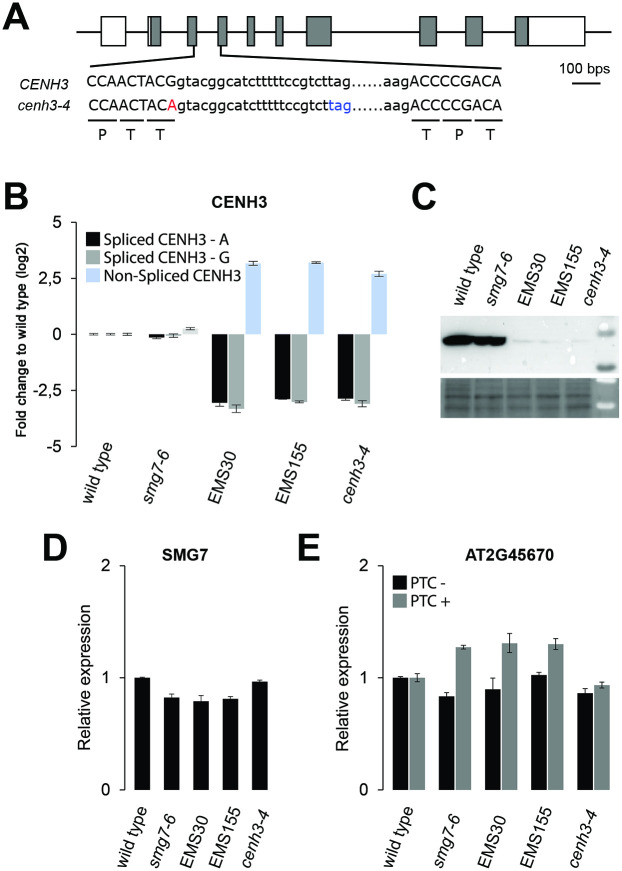
Fig 6. Molecular characterization of the cenh3-4 allele.
(A) Diagram of the CENH3 gene with exons marked as boxes. DNA sequence surrounding the intron 3 is shown; capital letters depict exons. Amino acids encoded by canonically spliced mRNA are indicated. The cenh3-4 mutation at the splice donor site of exon 3 is indicated in red, stop codon in the intron of unspliced mRNA in blue. (B) Relative abundance of spliced and unspliced CENH3 mRNA determined by quantitative RT-PCR. Two sets of primers were designed for fully spliced mRNA matching either wild type (CENH3-G) or cenh3-4 (CENH3-A) allele sequences. (C) Western blot detection of total CENH3 protein by anti-CENH3 antibody. Protein loading is shown on a Ponceau-S stained membrane (bottom panel). (D) Abundance of SMG7 mRNA relative to wild type determined by qRT-PCR. (E) NMD efficiency assessed by qRT-PCR analysis of alternatively spliced variants of the AT2G45670 gene with or without a premature termination codon (PTC) relative to wild type. Error bars in (B), (D) ad (E) represent standard deviations from 3 biological replicas.

