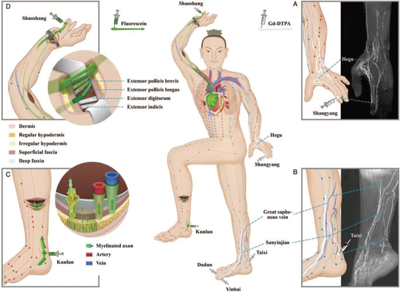
Figure 1.
A diagram of the acupoint-originated ISF flow pathways in the human body. Red, green dots and lines on the body surface represent the acupoints and main meridians in the atlas currently used in TCM. (A, B) shows the contrast-enhanced ISF flow pathways (white lines) originating from the acupoints in hand or foot by MRI. (C) shows the fluorescent ISF flow pathways (green arrows) from an acupoint in an ex vivo human leg sample, who took the acupoint injection before the lower leg amputation due to his severe foot gangrene. (D) shows the fluorescent ISF flow pathways from an acupoint in a cadaver that was performed by repeated chest compressions for 2.5 hours after the injection. (A) The ISF flow pathways (white lines) originating from Shangyang (the Jing-Well acupoint of Large Intestine Meridian) of a volunteer were found converging into one intersection point or pass by the downstream acupoint of Hegu in dorsal hand. (B) Originated from the three upstream acupoints of Yinbai in Spleen Meridian, Dadun in Liver Meridian, and Taixi in Kidney Meridian, it was found that the three ISF pathways (white lines) pass by the downstream acupoint of Sanyinjiao in different depths instead of converging into one intersection point. (C) At least four types of the fluorescent ISF pathways (green arrows) originating from Kunlun (the Jing-River point of the Urinary Bladder Meridian) were found in lower leg, including the cutaneous, perivenous, periarterial, and neural pathways. (D) The fluorescent ISF pathways (green arrows) originating from Shaoshang (the Jing-Well point of the Lung Meridian) were found to flow along the upper arm toward the right atrium and pericardium via the cutaneous pathway and venous PACT pathway along the veins in arm, superior vena cava, into the right atrial wall. The cutaneous pathways from the thumb can be seen on the skin of hand and lower forearm but not the skin above the level of the cubital fossa. When the skin of the wrist was opened layer by layer, the cutaneous pathways were found to contain layers of connective tissues, including the dermic, hypodermic, superficial, and deep fascial tissues on the tendons associated with thumb but not the tendons from the index finger. ISF: Interstitial fluid; MRI: Magnetic resonance imaging; PACT: Perivascular and adventitial connective tissue; TCM: Traditional Chinese medicine.

