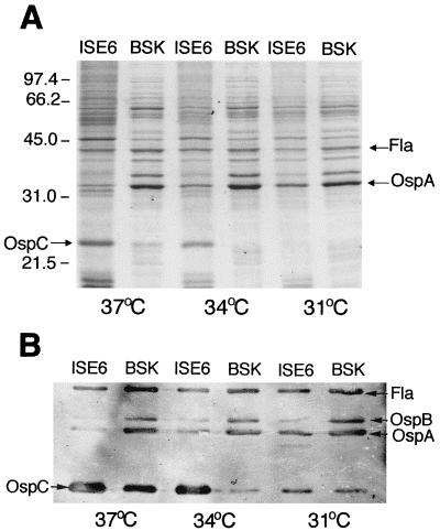
FIG. 3.
Analysis of B. burgdorferi JMNT by SDS-PAGE with Rapid Coomassie blue staining (A) or immunoblotting (B) with MAbs to OspA, OspB, OspC, and flagellin as probes. Spirochetes were cocultivated with a vector tick cell line (ISE6 cells) or were grown axenically in BSK medium at 37, 34, or 31°C. Molecular mass markers (in kilodaltons) are shown on the left.

