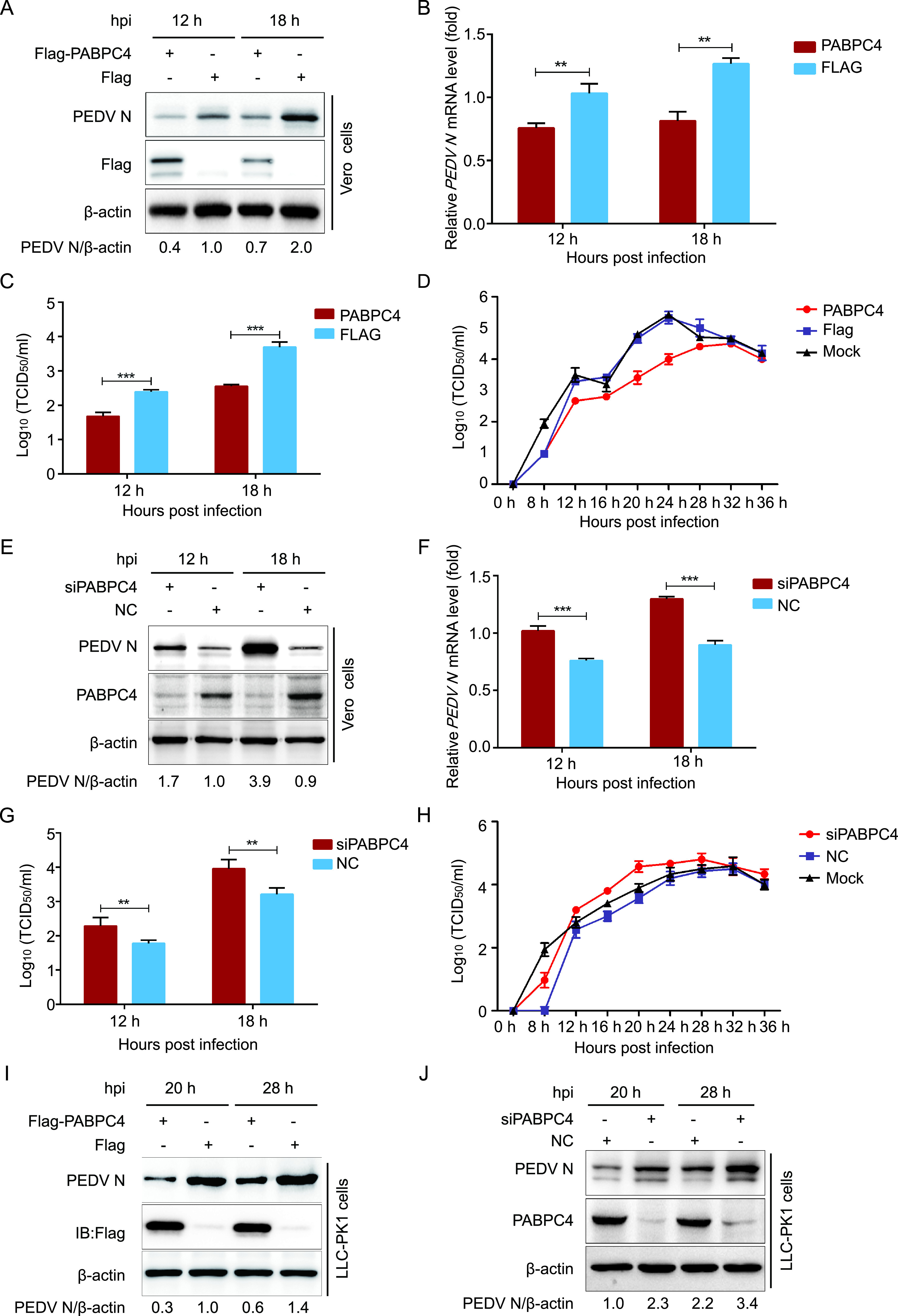
FIG 2.

PABPC4 inhibits PEDV replication. (A) Vero cells were transfected with plasmid encoding FLAG-PABPC4 or the FLAG tag. The cells were infected with PEDV at an MOI of 0.01 after 24 h posttransfection and harvested at the indicated times. The N protein was analyzed with Western blotting. β-actin was used as the sample loading control. (B) The PEDV N mRNA in the same samples (A) were analyzed by real-time PCR. (C and D) PEDV titers in the culture supernatants of the Vero cells treated as described in panel A were measured as 50% tissue culture infective dose (TCID50). (E) PABPC4 siRNA or negative control siRNA were transfected into Vero cells, and then infected with PEDV at an MOI of 0.01. The N protein was analyzed with Western blotting. β-actin was used as the sample loading control. (F) The PEDV N mRNA in the same samples (E) were analyzed by real-time PCR. (G and H) PEDV titers in the culture supernatants of the Vero cells treated as described in panel E were measured as TCID50. (I and J) FLAG-PABPC4 vector or PABPC4 siRNA were transfected into LLC-PK1 cells, and then infected with PEDV at an MOI of 1. The N protein was analyzed with Western blotting. β-actin was used as the sample loading control. Data are represented as means ± SD of triplicate samples. **, P < 0.01; ***, P < 0.001 (two-tailed Student’s t test).
