Figure 5.

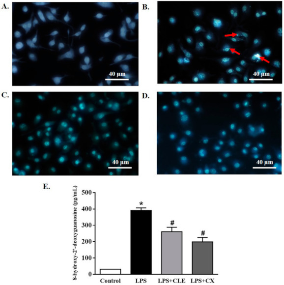
The effects of CLE on DNA damage in LPS-stimulated RAW 264.7 cells. Cells were plated in 12-well plates and treated with CLE or CX in the presence or absence of LPS. Cells were treated 24 h later with Hoechst 33,342 staining at 5 µg/mL for 10 min and then observed under an inverted fluorescence microscope (original magnification, ×40). The change in the nucleus of apoptotic cells is shown by the arrows (A–D). The amount of 8-OHdG in the DNA was determined using an 8-OHdG-EIA kit (E). Values shown are mean ± SEM (n = 5); * p < 0.05 vs. control and # p < 0.05 vs. LPS.
