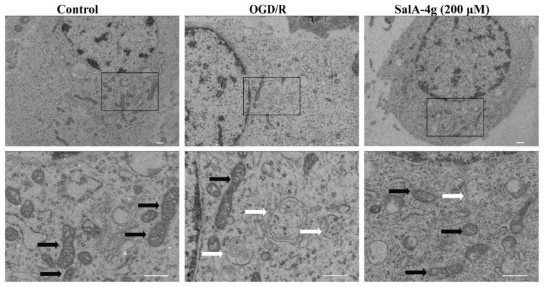
Figure 3.
SalA-4g stabilizes the mitochondrial structure in cortical neurons under OGD/R stress. Representative TEM images of control cells, cells exposed to 2.5 h of OGD and 24 h of reoxygenation alone, and cells treated with 200 μM SalA-4g during OGD/R period. White arrows indicate damaged mitochondria and black arrows indicate healthy mitochondria. Scale bar, 0.5 μm.

