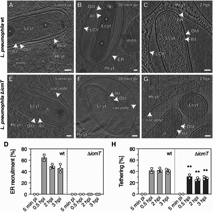
FIG 1.
L. pneumophila wild-type tethers its cell pole to the LCV membrane at early and late infection stages. A. castellanii amoebae were infected with L. pneumophila wild-type (A to C) or the ΔicmT mutant (E to G) for the time indicated. Representative 2D images of cryotomograms (12-nm tomographic slices) of L. pneumophila wild-type residing in a tight phagosome at very early infection stages (5 min pi, nvacuoles = 10) (A) or in a mature, ER-decorated LCV (30 min pi, nLCVs = 29; 2 h pi, nLCVs = 38) (B, C), with the cell pole tethered to the LCV membrane (∼41% on average over time, ncell poles = 202). (D) Quantification of data shown in panels A to C and E to G. (E to G) T4SS-defective ΔicmT mutant bacteria oriented their poles to the vacuole membrane less frequently (∼28% on average over time, ncell poles = 45). (H) Quantification of data shown in panels A to C and E to G. Data in panels D and H are represented as mean ± standard deviation (SD) from at least two independent infection experiments (one-way ANOVA test; **, P < 0.01). OM, outer membrane; IM, inner membrane; Pha, phagosome; LCV, LCV membrane; Lcyt, L. pneumophila cytoplasm; Hcyt, host cell cytoplasm; ER, endoplasmic reticulum; mito, mitochondrion; asterisk, storage granule; Scale bars, 100 nm.

