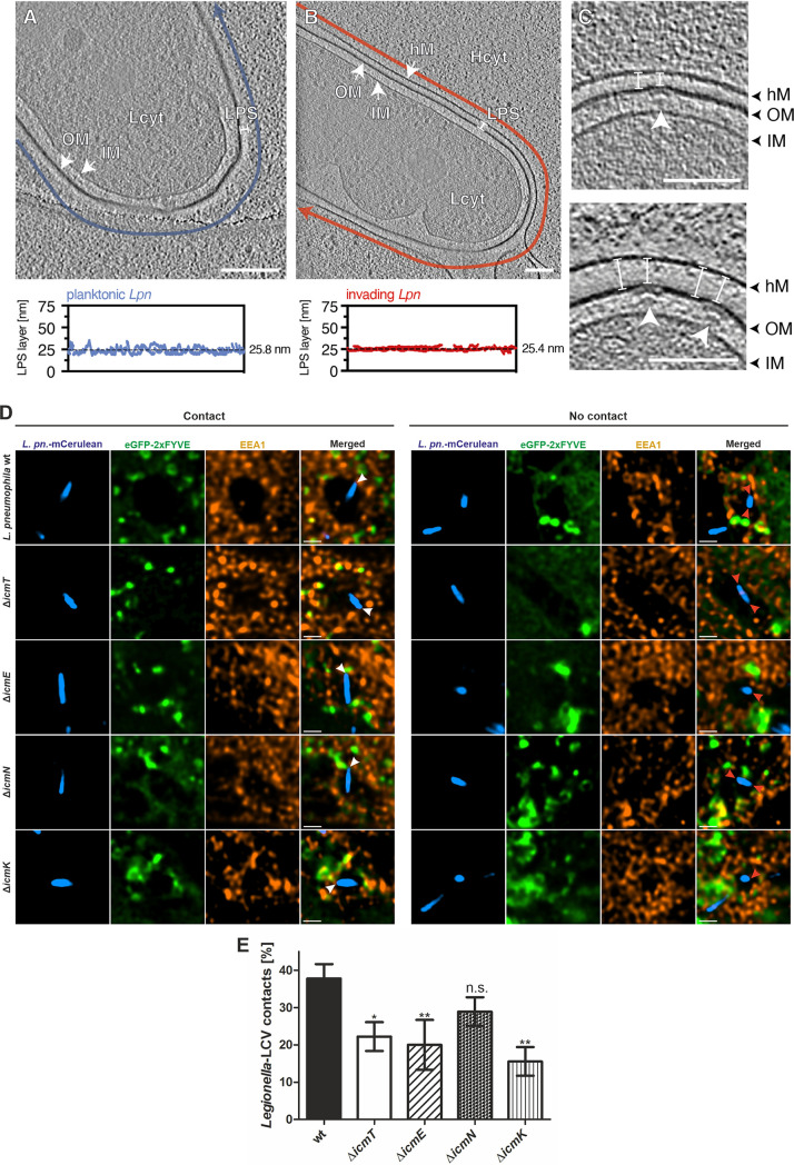
FIG 5.
LPS layers at Icm/Dot sites and L. pneumophila poles tethered to host membranes. L. pneumophila LPS layer (A, B) or Icm/Dot T4SS (C) in planktonic bacteria (A) or infected (MOI of 75, 5 min pi) HeLa cells (B, C). The LPS layer thickness in planktonic L. pneumophila (∼26 nm; blue) is comparable to the gap between the bacterium and the HeLa cell (∼25 nm; red), suggesting that LPS serves as a physical barrier during host cell invasion. The distance between the bacterial outer membrane and the host cell membrane was constant along the bacterial cell body, indicated by a low standard deviation (B, bottom; approximately ±2 nm). Planktonic and adherent L. pneumophila show similar LPS layer thickness. (C) At T4SS assembly sites, the gap between the bacterium and the HeLa cell is slightly reduced (∼19 nm versus ∼25 nm, white arrowhead). Shown are 8-nm tomographic slices of cryotomograms. One tomogram was used for the quantification of the LPS layer as a representative of the population. Blue and red arrows indicate the path along which the thickness of the LPS layer was quantified. OM, outer membrane; IM, inner membrane; hM, host cell membrane; Lcyt, L. pneumophila cytoplasm; Hcyt, host cell cytoplasm; LPS, lipopolysaccharide; white arrowhead, T4SS; Scale bars, 100 nm. (D) Representative fluorescence micrographs of HeLa cells producing eGFP-2×FYVE (pEGFP-2×FYVE) and infected (MOI of 150; 1 h) with mCerulean-producing L. pneumophila wild-type or ΔicmT, ΔicmE, ΔicmN, or ΔicmK mutant bacteria harboring plasmid pNP99. Infected cells were fixed with PFA and stained with an anti-early endosome antigen 1 (EEA1) antibody prior to imaging. Examples are shown for contact between bacteria and the LCV membrane (left, white arrowheads) or no contact (right, red arrowheads). Scale bars, 1 μm. (E) Quantification of data shown in panel D (nevents = 45). Data are represented as mean ± SD from three biological replicates (one-way ANOVA test; *, P < 0.05; **, P < 0.01; n.s., not significant).

