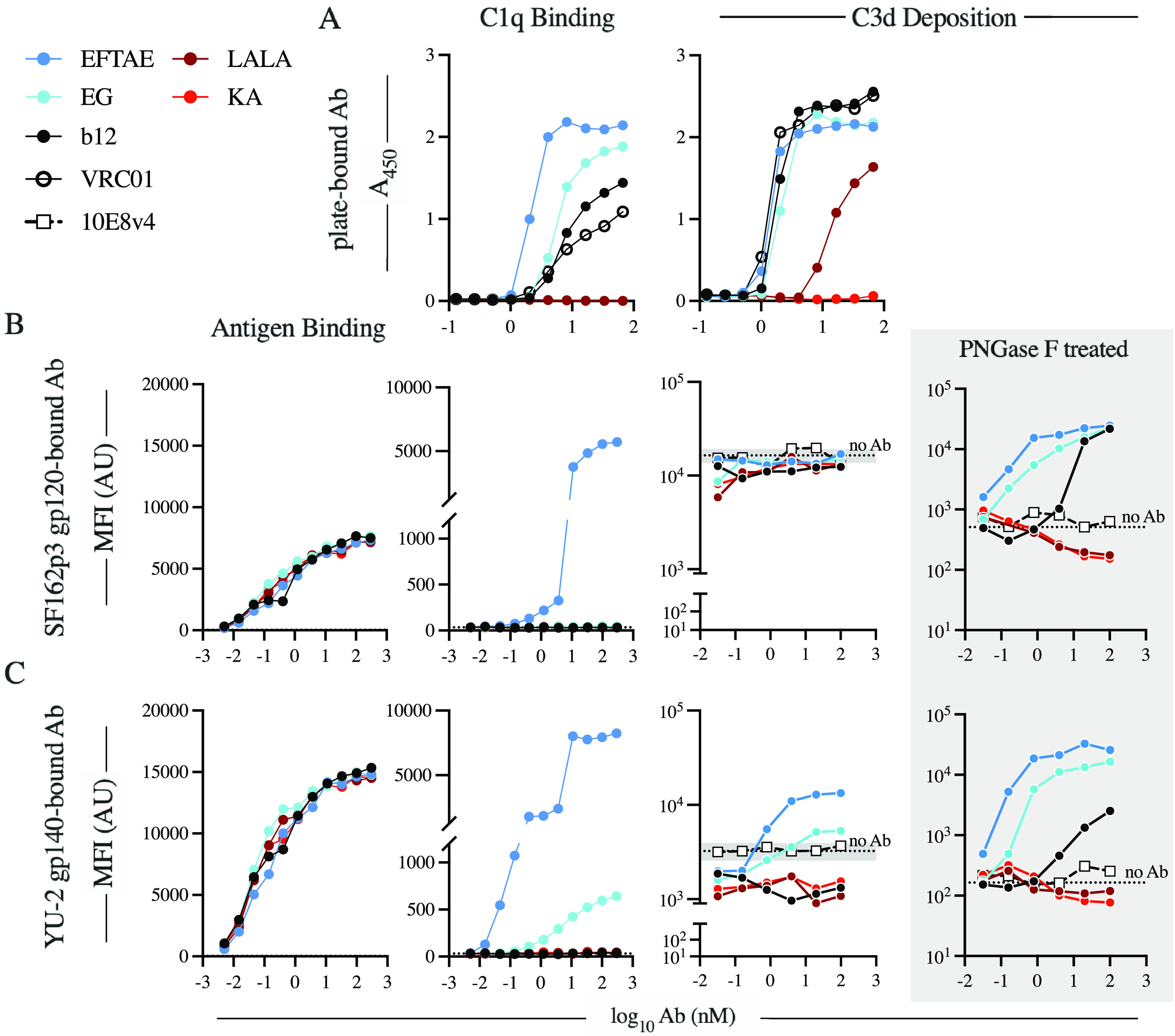
FIG 2.

The ability of b12 to activate complement is influenced by assay setup and antigen context. (A) The antigen-independent ability of the antibody panel to bind C1q (left) and fix complement (right) was determined by ELISAs via antibody-coated wells. (B and C) Beads conjugated with SHIVSF162P3 gp120 (B) or HIV-1YU-2 gp140 trimer (C) were used to assay antigen binding (left), C1q binding (center), and complement fragment C3d deposition (right). Antigen beads treated with PNGase F were used to assess the impact of N-linked antigen glycosylation antibody-independent activation and to isolate antibody-dependent C3d deposition (shaded). For C3d deposition on non-PNGase F-treated antigen beads, background activity is reported as the average MFI (dotted line) ± standard deviation (shaded region on the y axis) of anti-C3d detected on beads in replicate wells of pooled NHS (n = 6) in the absence of antibody. Data are representative of results from two independent experiments. AU, arbitrary units.
