
Figure 4.
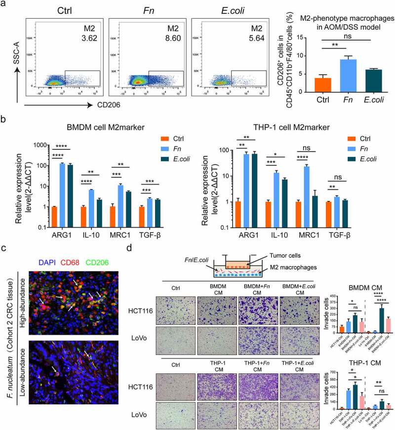
F. nucleatum induced the M2 polarization of macrophage to promote CRC metastasis. (a) Frequencies of M2-phenotype macrophage (CD45+ CD11b+ F4/80+ CD206+) from colon lamina propria in AOM/DSS model after F. nucleatum or PBS or E. coli treatment were tested by multicolor flow cytometry. (Student’s t test). (b)The mRNA expression of M2 markers (ARG1, MRC1, IL-10 and TGF-β) were detected in THP-1-derived macrophage and BMDMs after being infected with F. nucleatum or E. coli or PBS for 24 h (Student’s t test). (c) Representative images of M2-phenotype macrophage (CD68+ CD206+) were examined by immunofluorescence in F. nucleatum high and low abundance CRC tissues. Scale bars:25 μm. The white arrows indicated CD68+ CD206+ M2-phenotype macrophage. (d) After co-cultured with culture medium of Macrophage pretreated with F. nucleatum or E. coli or PBS for 24 h, the mobility of CRC cells was detected by transwell assay (Each experiment was repeated in triplicate; Student’s t test). Scale bars:100 μm. * p < .05, ** p < .01, and *** p < .001, **** p < .0001, ns no significant